
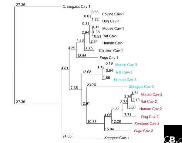
The caveolingenefamily has three members in vertebrates: caveolin-1, caveolin-2, and caveolin-3. So far, most caveolin-related analysis has been carried out in mammals, however the proteins have additionally been present in different animals, together with Xenopus laevis, Fugu rubripes, and Caenorhabditis elegans.
Caveolins can function protein markers of caveolae (‘little caves’), invaginations within the plasma membrane 50-100 nanometers in diameter. Caveolins are discovered predominantly on the plasma membrane but additionally within the Golgi, the endoplasmic reticulum, in vesicles, and at cytosolic areas. They are expressed ubiquitously in mammals, however their expression ranges range significantly between tissues.
The highest ranges of caveolin-1 (additionally referred to as caveolin, Cav-1 and VIP2I) are present in terminally-differentiated cell sorts, corresponding to adipocytes, endothelia, easy muscle cells, and sort I pneumocytes. Caveolin-2 (Cav-2) is colocalized and coexpressed with Cav-1 and requires Cav-1 for correct membrane concentrating on; the Cav-2 gene additionally maps to the identical chromosomal area as Cav-1 (7q31.1 in people). Caveolin-3 (Cav-3) has better protein-sequence similarity to Cav-1 than to Cav-2, however it’s expressed primarily in muscle cells, together with easy, skeletal, and cardiac myocytes. Caveolins take part in lots of necessary mobile processes, together with vesicular transport, ldl cholesterol homeostasis, sign transduction, and tumor suppression.
Limb-girdle muscular dystrophy (LGMD) is a clinically and genetically heterogeneous group of myopathies, together with autosomal dominant and recessive types. To date, two autosomal dominant types have been acknowledged: LGMD1A, linked to chromosome 5q, and LGMD1B, related to cardiac defects and linked to chromosome 1q11-21.
Here we describe eight sufferers from two totally different households with a brand new type of autosomal dominant LGMD, which we suggest to name LGMD1C, related to a extreme deficiency of caveolin-Three in muscle fibres. Caveolin-3 (or M-caveolin) is the muscle-specific type of the caveolin protein household, which additionally consists of caveolin-1 and -2. Caveolins are the principal protein parts of caveolae (50-100 nm invaginations present in most cell sorts) which symbolize appendages or sub-compartments of plasma membranes.
We localized the human caveolin-3 gene (CAV3) to chromosome 3p25 and recognized two mutations within the gene: a missense mutation within the membrane-spanning area and a micro-deletion within the scaffolding area. These mutations might intervene with caveolin-Three oligomerization and disrupt caveolae formation on the muscle cell plasma membrane.
Caveolae are specialised invaginations of the plasma membrane present in quite a few cell sorts. They have been implicated as enjoying a task in a wide range of physiological processes and are sometimes characterised by their affiliation with the caveolinfamily of proteins.
We present right here by the use of focused gene disruption in mice {that a} distinct caveolae-associated protein, Cavin/PTRF, is a vital part of caveolae. Animals missing Cavin haven’t any morphologically detectable caveolae in any cell sort examined and have markedly diminished protein expression of all three caveolin isoforms whereas retaining regular or above regular caveolin mRNA expression.
Cavin-knockout mice are viable and of regular weight however have larger circulating triglyceride ranges, considerably decreased adipose tissue mass, glucose intolerance, and hyperinsulinemia–characteristics that represent a lipodystrophic phenotype. Our outcomes underscore the multiorgan function of caveolae in metabolic regulation and the obligate presence of Cavin for caveolae formation.
Bruton’s tyrosine kinase (Btk): operate, regulation, and transformation with particular emphasis on the PH area
Bruton’s agammaglobulinemia tyrosine kinase (Btk) is a cytoplasmic tyrosine kinase necessary in B-lymphocyte improvement, differentiation, and signaling.
Btk is a member of the Tec household of kinases. Mutations within the Btk gene result in X-linked agammaglobulinemia (XLA) in people and X-linked immunodeficiency (Xid) in mice. Activation of Btk triggers a cascade of signaling occasions that culminates within the generation of calcium mobilization and fluxes, cytoskeletal rearrangements, and transcriptional regulation involving nuclear factor-kappaB (NF-kappaB) and nuclear issue of activated T cells (NFAT).
In B cells, NF-kappaB was proven to bind to the Btk promoter and induce transcription, whereas the B-cell receptor-dependent NF-kappaB signaling pathway requires useful Btk. Moreover, Btk activation is tightly regulated by a plethora of different signaling proteins together with protein kinase C (PKC), Sab/SH3BP5, and caveolin-1.
For instance, the prolyl isomerase Pin1 negatively regulates Btk by lowering tyrosine phosphorylation and regular state ranges of Btk. It is intriguing that PKC and Pin1, each of that are damaging regulators, bind to the pleckstrin homology area of Btk. To this finish, we describe right here novel mutations within the pleckstrin homology area investigated for his or her reworking capability. In explicit, we present that the mutant D43R behaves much like E41Ok, already identified to own such exercise.